

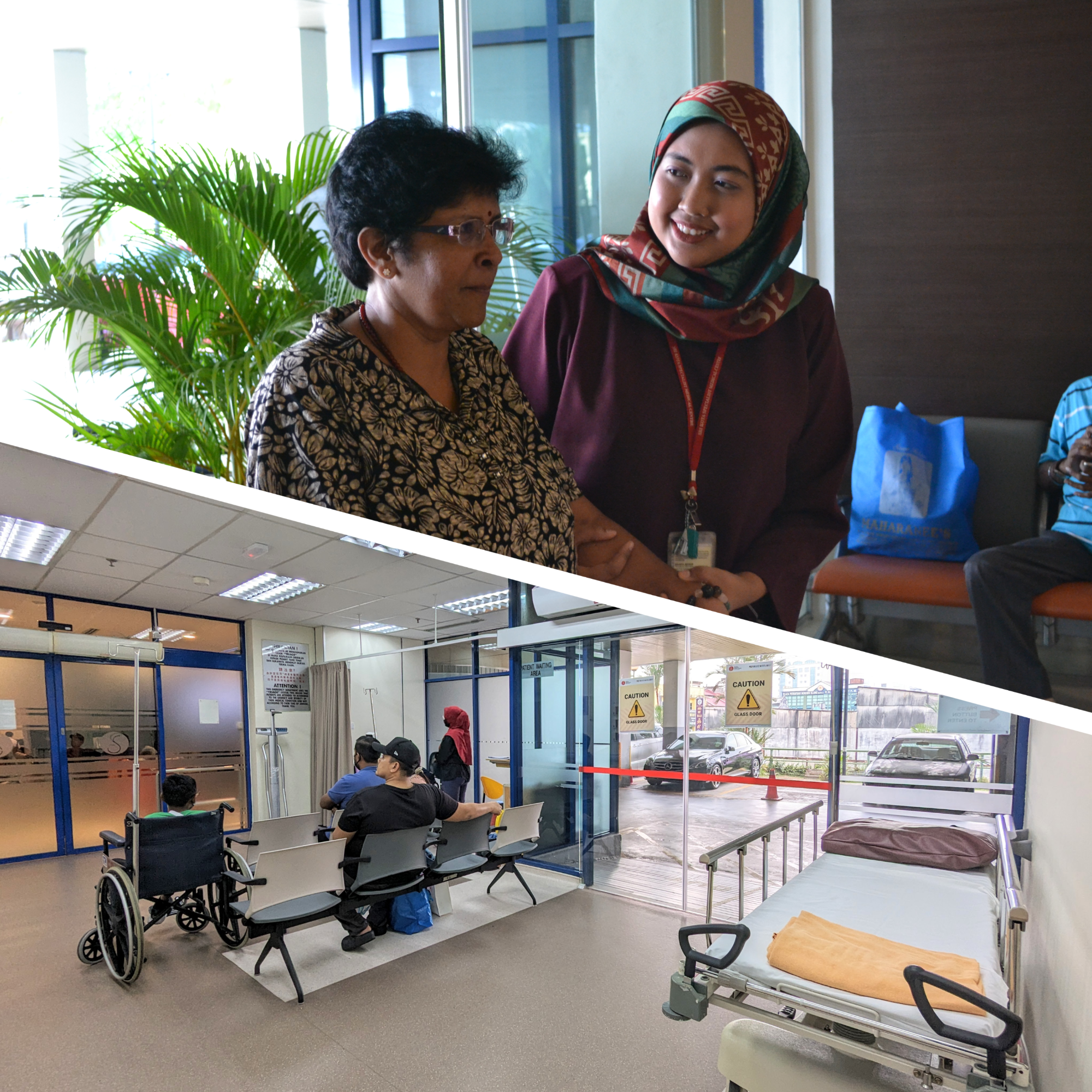
Welcome to the Sri Kota Stroke Centre – An award-winning Stroke Centre that offers 24-hour diagnosis, treatment, and rehabilitation of stroke patients. Our team of experts is dedicated to providing the highest level of care for patients who have suffered a stroke. Our state-of-the-art facility and experienced staff ensure that patients receive rapid and effective treatment, leading to better outcomes and a faster recovery. Within 60 minutes of arrival at the Accident & Emergency Department, patients are assured that they are assessed, undergo a head scan, scan interpretation and treatment decisions of clot-dissolving medicines are made.
Services Offered:
- Rapid diagnosis and treatment of stroke
- 24/7 emergency care for stroke patients
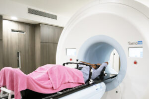
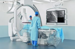
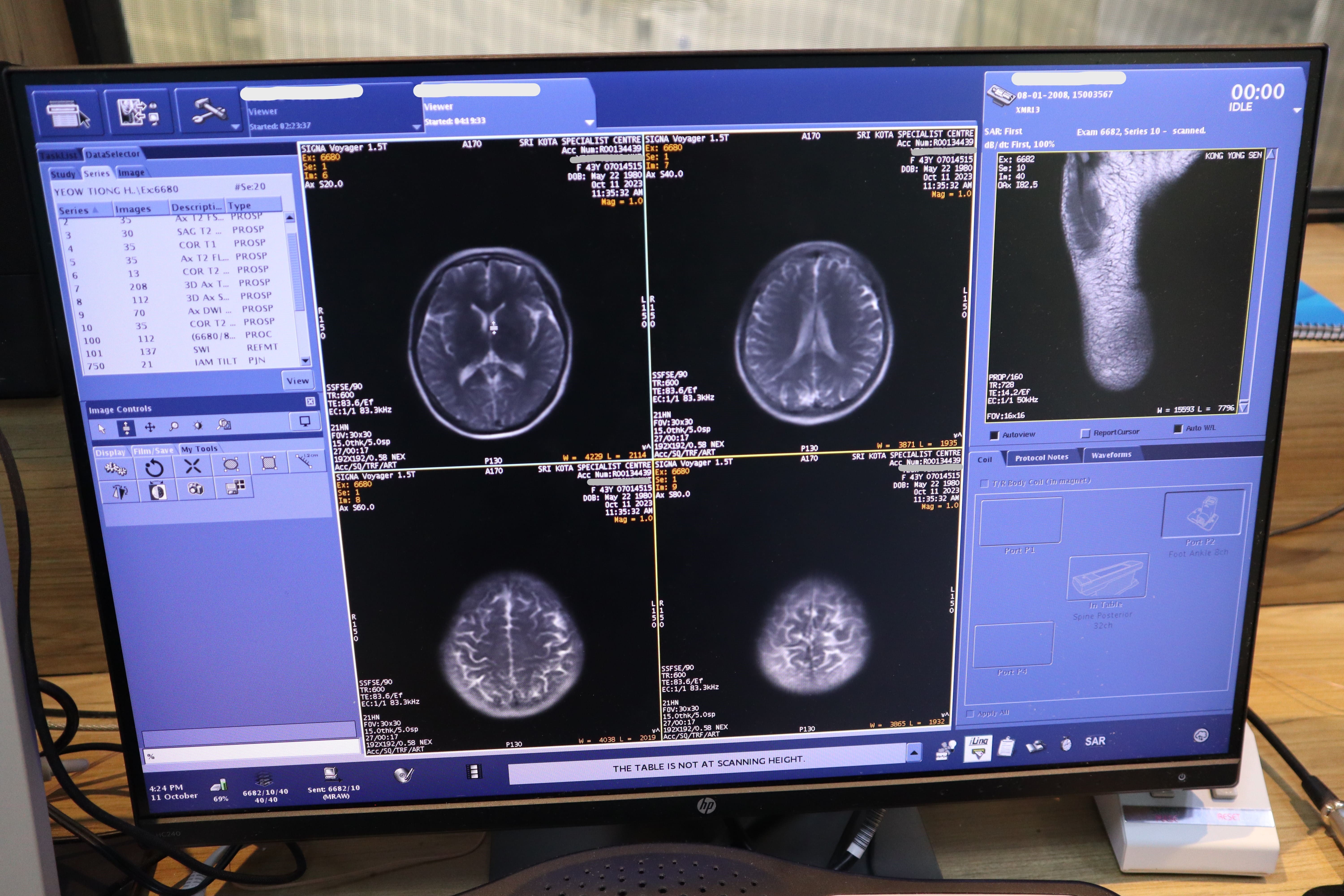
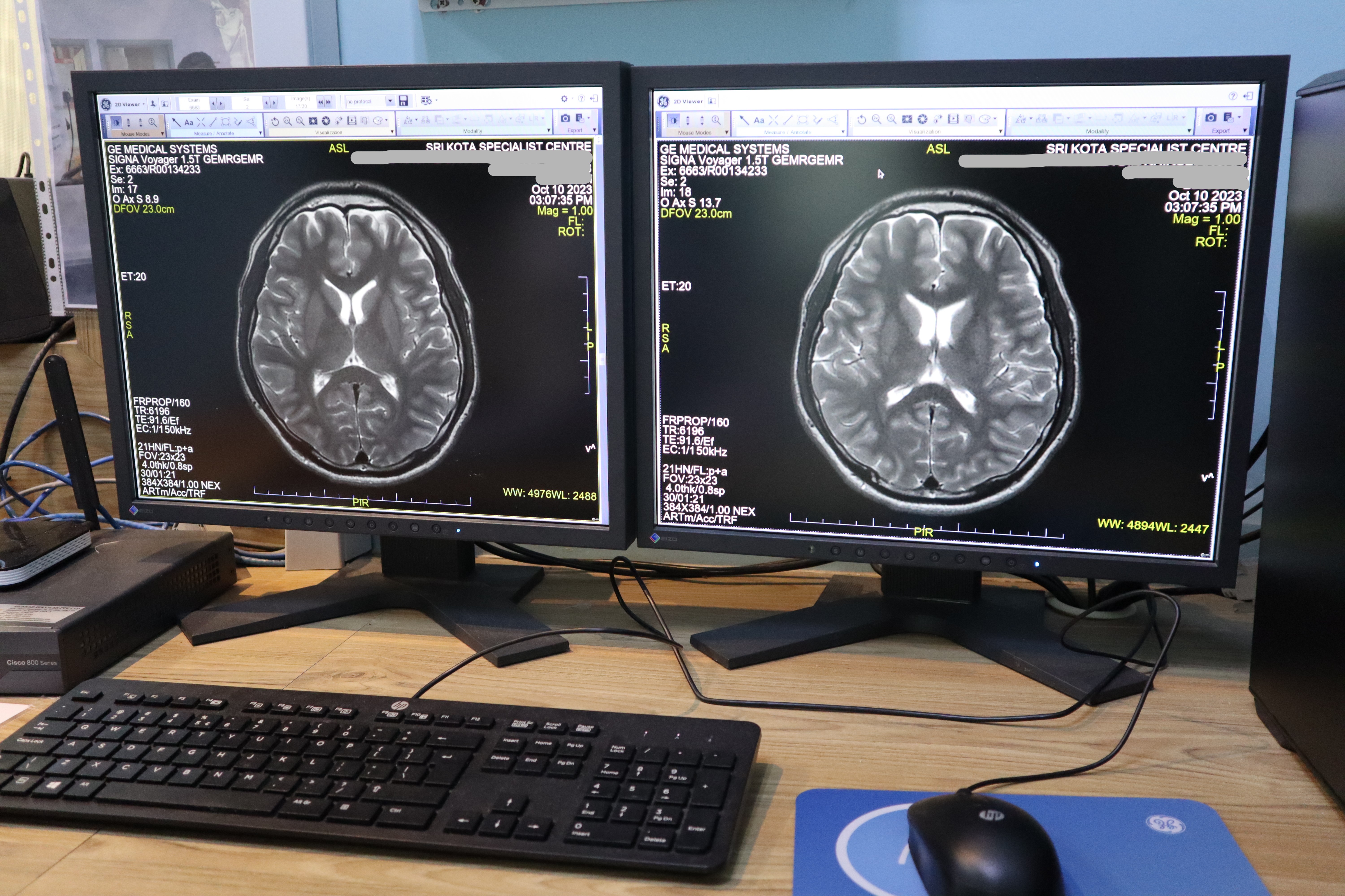
- Cutting-edge technology and equipment, including CT and MRI scans
- Comprehensive stroke rehabilitation services
- On-site specialists in neurology, neurosurgery, and rehabilitation medicine
- Patient-centred care focused on individual needs and goals
Diagnosis and Treatment:
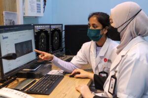
At our stroke centre, we prioritize rapid diagnosis and treatment to minimize the damage caused by stroke. Our team of specialists works closely with the Emergency Department to ensure that patients receive prompt attention upon arrival. We use the latest technology and equipment, including CT and MRI scans, to quickly and accurately diagnose the type and severity of stroke. Our team of experts then develops a personalized treatment plan that may include medication, surgery, or other interventions.
Rehabilitation:
Stroke can cause a wide range of physical and cognitive impairments, but rehabilitation can help patients regain function and improve their quality of life. Our stroke centre has a dedicated team of rehabilitation specialists who work with patients to develop personalized treatment plans. Our approach is patient-centred, meaning that we focus on individual needs and goals. Our rehabilitation services include physical therapy, occupational therapy, speech therapy, and more. Our comprehensive stroke centre in Malaysia provides holistic care for stroke survivors, aiding them in their journey to recovery.
Specialists:
Our stroke centre has on-site specialists in neurology, neurosurgery, and rehabilitation medicine. This means that our patients receive comprehensive care from a team of experts who are experienced in treating stroke patients. Our specialists work closely together to ensure that patients receive the best possible care at all stages of treatment and rehabilitation.
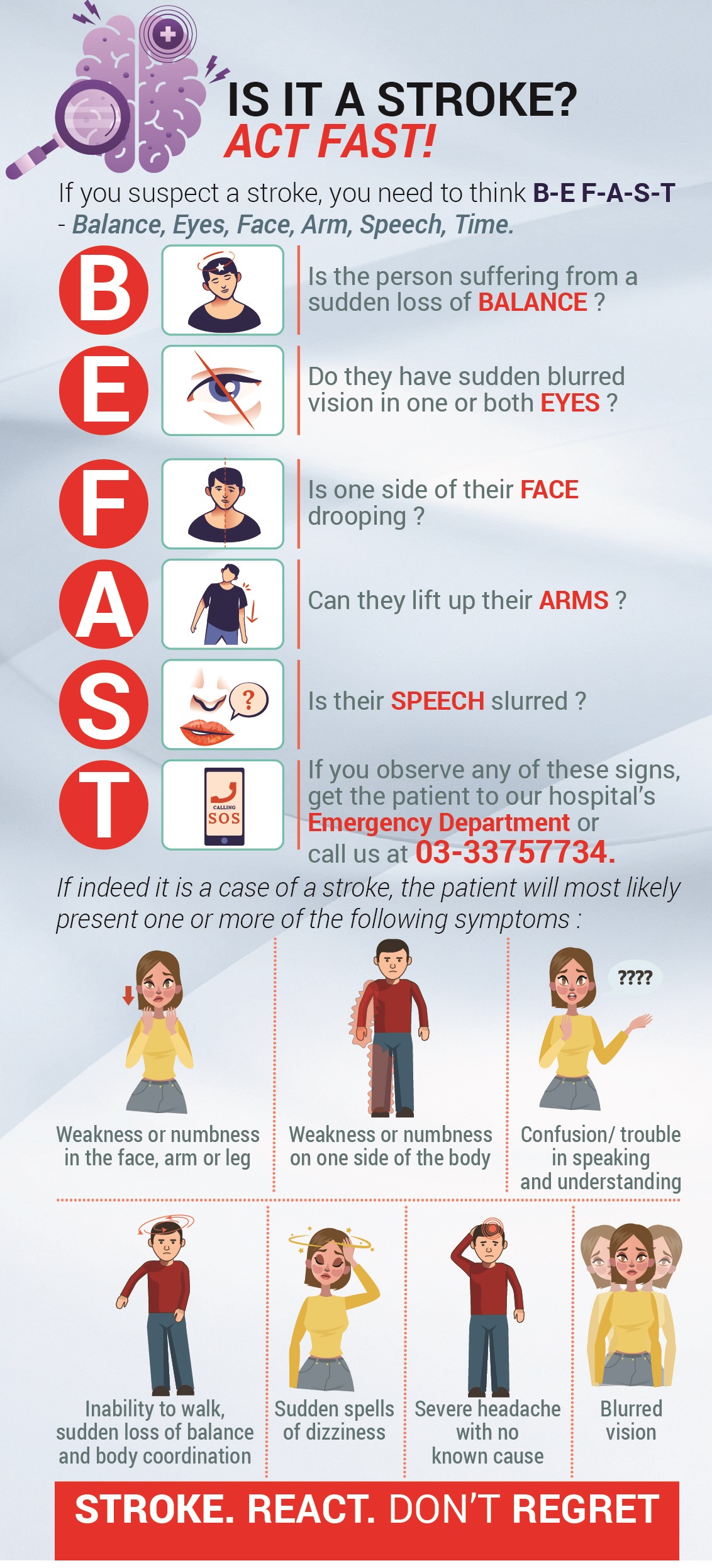
If you or someone you know is experiencing symptoms of stroke, it is important to seek medical attention immediately. Time is of the essence in stroke treatment, and the faster a stroke is diagnosed and treated, the better the chances of a good outcome. Our stroke centre is available 24/7 to provide emergency care for stroke patients.
Our Multidisciplinary team consists of:
- Consultant Neurologist
- Consultant Neurosurgeon
- Consultant Emergency Physician
- Consultant Cardiologist
- Vascular and Endovascular Surgeon
- Physiotherapist
- Dietitian
Contact us with Direct2Doctor Careline at 03-3375 7734 to learn more about our services and how we can help you or your loved one recover from a stroke.

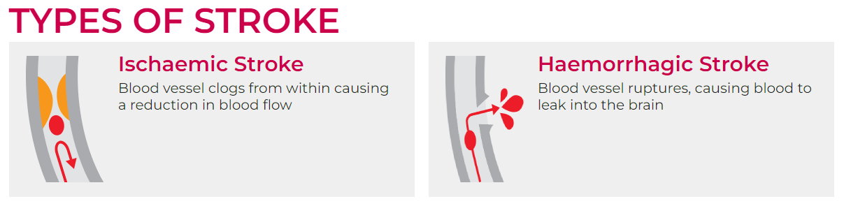
Ischemic Stroke:
- Ischemic strokes are the most common type, accounting for about 87% of all strokes.
- They occur when a blood vessel that supplies blood to the brain is blocked by a clot (thrombus) or embolism. This blockage restricts blood flow and oxygen to a specific part of the brain.
- Subtypes of ischemic strokes include:
a. Thrombotic Stroke: Caused by a blood clot (thrombus) that forms within an artery supplying the brain.
b. Embolic Stroke: Occurs when a clot (embolus) travels from another part of the body and blocks a brain artery.
c. Cryptogenic Stroke: When the cause of the stroke is unclear.
Hemorrhagic Stroke:
- Hemorrhagic strokes are less common but often more severe than ischemic strokes. They occur when a blood vessel in the brain ruptures, causing bleeding into the surrounding tissue.
- Subtypes of hemorrhagic strokes include:
a. Intracerebral Hemorrhage: Bleeding directly into brain tissue, usually caused by the rupture of small blood vessels.
b. Subarachnoid Hemorrhage: Bleeding into the space between the brain and the surrounding membrane (subarachnoid space), often caused by a ruptured aneurysm.
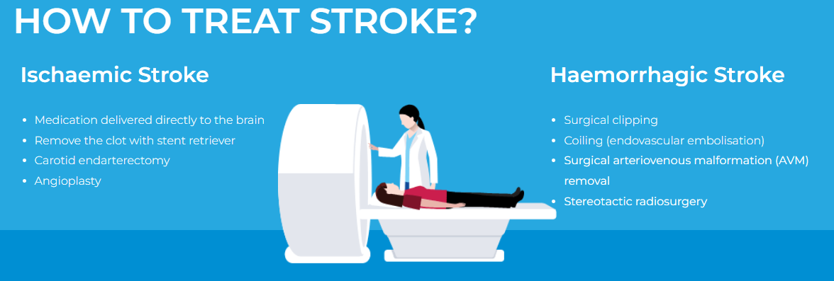
image source: https://mystrokehospital.my/what-is-stroke/
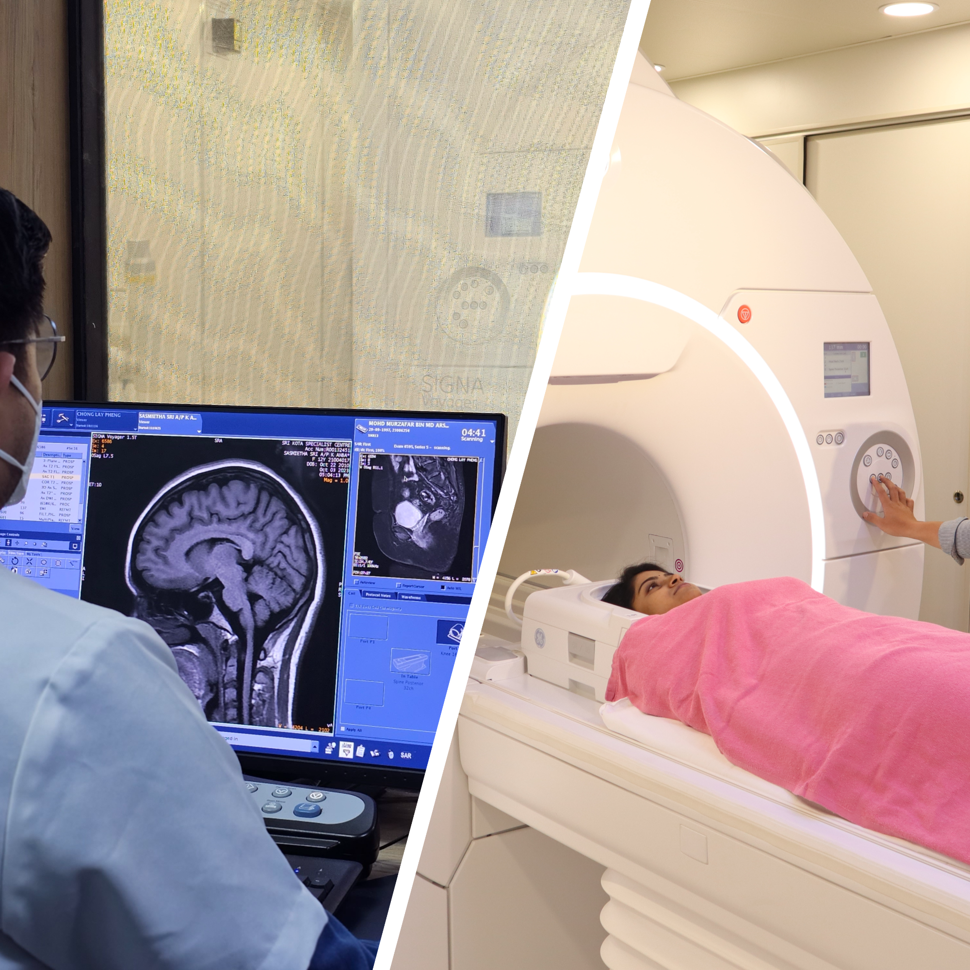
In the context of stroke, both brain MRI (Magnetic Resonance Imaging) and brain CT (Computed Tomography) have distinct roles and characteristics when it comes to the evaluation and diagnosis of stroke. Here’s how they differ in this context:
- Time Sensitivity:
- Brain MRI: MRI is more sensitive to early changes in brain tissue. It can detect ischemic stroke (caused by a blood clot or blocked blood vessel) within minutes to hours of onset. It can also distinguish between acute and chronic strokes.
- Brain CT: CT is often the initial choice for stroke assessment, especially in the acute phase, as it can quickly rule out hemorrhagic stroke (caused by bleeding in the brain), which may require immediate surgical intervention. CT is more readily available in emergency settings and can provide a rapid assessment of acute intracranial bleeding.
- Detection of Stroke Type:
- Brain MRI: MRI is better at differentiating between ischemic and hemorrhagic strokes and can provide more information about the extent of brain damage associated with ischemic strokes. It can help identify the specific regions of the brain affected by the stroke and assess the age of the stroke (acute, subacute, or chronic).
- Brain CT: CT is more efficient at identifying the presence of hemorrhagic strokes and can quickly determine if there is bleeding in the brain. However, it is less precise in distinguishing between different types of ischemic strokes.
- Tissue Characterization:
- Brain MRI: MRI provides detailed information about the brain’s soft tissues, making it valuable for assessing the extent of damage to brain tissue surrounding the stroke and for identifying areas of potential salvage.
- Brain CT: CT provides a more limited view of soft tissues, and while it can identify hemorrhage, it may not reveal subtle changes in the brain tissue caused by ischemic stroke in its early stages.
- Thrombolytic Therapy Decisions:
- Brain MRI: MRI can help in identifying patients who may benefit from thrombolytic therapy (clot-dissolving medication) and in determining the optimal treatment window, as it can visualize the ischemic penumbra (potentially salvageable brain tissue) and differentiate it from the irreversibly damaged tissue.
- Brain CT: CT is often used to quickly determine if hemorrhagic stroke is present, as thrombolytic therapy is contraindicated in such cases. However, CT alone may not provide the same level of information about the ischemic penumbra as MRI.
- Follow-Up and Monitoring:
- Brain MRI: MRI is often used for follow-up imaging after a stroke to assess recovery, track changes in brain tissue, and evaluate the effectiveness of treatment.
- Brain CT: CT may be used for initial assessment and monitoring in cases where rapid follow-up is needed, but it may be complemented by MRI for more detailed assessments later on.
In summary, both brain MRI and brain CT play important roles in the evaluation of stroke, with CT often serving as the initial tool to quickly rule out hemorrhagic stroke, while MRI is used for more detailed assessment, especially in the context of ischemic stroke, its subtypes, and follow-up evaluations. The choice between the two depends on the clinical scenario, time constraints, and the specific information needed for treatment decisions.